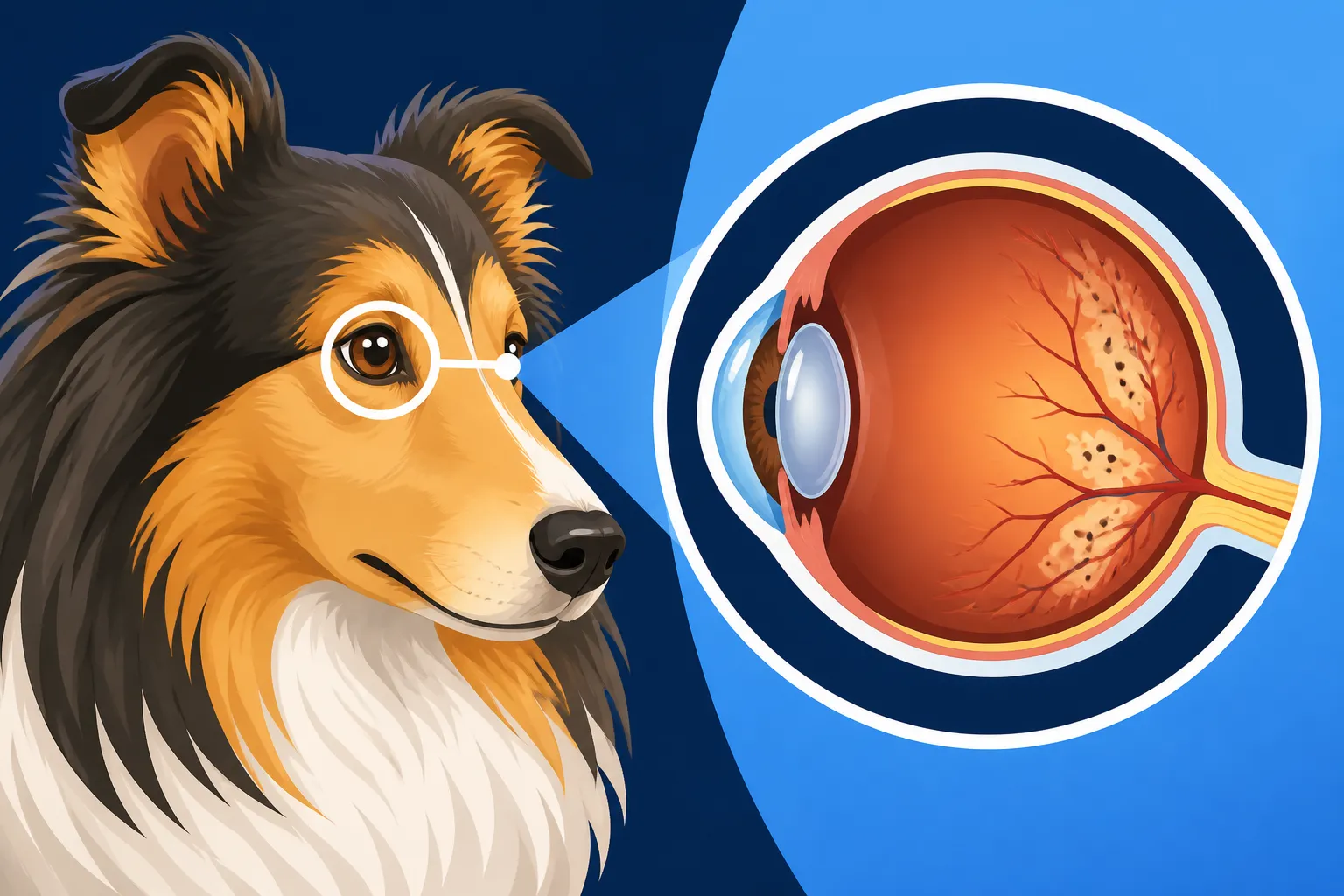
What Collie Eye Anomaly Does to the Back of the Eye
It’s truly disheartening when you learn that your beloved Collie, a breed known for its stunning beauty and intelligent eyes, might be predisposed to a genetic condition that affects their vision. Collie Eye Anomaly (CEA) is one of those frustrating realities that many Collie owners face, and it often leaves us wondering, "What exactly is happening inside my dog's eye?" This comprehensive guide is here to demystify Collie Eye Anomaly, explaining in detail what this inherited condition does to the delicate structures at the back of your Collie's eye.
Understanding Collie Eye Anomaly (CEA): The Basics
Collie Eye Anomaly, often simply called CEA, is a congenital, inherited disorder that impacts the development of the eye in Collies and related breeds like Australian Shepherds, Border Collies, and Shetland Sheepdogs. It's not something that develops later in life due to injury or illness; it’s a structural defect present from birth, stemming from improper development of the eye during gestation. The severity can range dramatically, from almost imperceptible changes that don't affect vision at all, to severe abnormalities that can lead to blindness. As a responsible Collie owner, understanding the nuances of CEA is paramount, not just for your current dog but for future generations, given its genetic nature. It's a condition that truly speaks to the importance of transparent breeding practices and thorough health testing.
The Genetic Roots of CEA
At its core, CEA is a genetic disorder, specifically an autosomal recessive trait. What this means for us non-genetics experts is that a dog must inherit two copies of the defective gene—one from each parent—to be affected by CEA. Dogs with only one copy are considered carriers; they won't show symptoms themselves, but they can pass the gene on to their offspring. This is why you might hear about "CEA clear," "CEA carrier," and "CEA affected" statuses when breeders discuss their dogs. The gene responsible for CEA is known as NHEJ1. The ability to test for this specific gene has been a monumental step forward for the breed, allowing breeders to make informed decisions and work towards reducing the prevalence of CEA. However, it's also important to remember that genetics can sometimes be tricky; even with testing, vigilance remains key. The widespread nature of this gene is why many experts often state that it’s safe to assume your Collie has the MDR1 mutation as well, highlighting the prevalence of genetic health concerns in the breed.
How CEA Develops in the Womb
The abnormalities associated with CEA aren't things that just pop up one day. They originate during the early stages of fetal development, typically around the fifth to sixth week of gestation. This is a critical period when the various structures of the eye are forming. In Collies with CEA, there’s a failure of complete or normal development in certain tissues, particularly the choroid, sclera, and retina. The choroid is a layer of blood vessels and connective tissue between the retina and the sclera (the white outer layer of the eye) that nourishes the retina. When it doesn't form correctly, or if there are structural defects in the sclera itself, it sets the stage for the various manifestations of CEA. It’s like a blueprint error in building a house; the foundation (the eye's early development) has flaws, and those flaws will impact the structural integrity of the finished product. This early developmental window is also why a definitive diagnosis can be made very early in a puppy's life.
Choroidal Hypoplasia: The Mildest Form of CEA
Choroidal hypoplasia, often abbreviated as CH, is generally considered the mildest and most common manifestation of Collie Eye Anomaly. In fact, when a veterinarian talks about a Collie being "affected" by CEA, they are most often referring to this particular form. Many Collies diagnosed with CH live full, happy lives with perfect vision, completely unaware they even have the condition. However, it's crucial not to dismiss it entirely, as even CH can sometimes mask or be accompanied by more severe issues, or predispose the eye to complications later on. Understanding CH helps us appreciate the spectrum of CEA and why careful eye exams are so important for puppies. For a deeper understanding of how widespread this issue is, it's worth exploring how many Collies are affected by Collie Eye Anomaly.
What Choroidal Hypoplasia Looks Like on Exam
During a thorough ophthalmic examination, a veterinary ophthalmologist will look for specific tell-tale signs of choroidal hypoplasia. The primary finding is a thinning or absence of the choroid, the vascular layer rich with blood vessels and pigment that lies beneath the retina. In a healthy eye, this layer gives the tapetum (the reflective layer behind the retina) its vibrant, iridescent sheen. With CH, the ophthalmologist will often observe a pale, often pinkish or translucent area, usually in the temporal quadrant of the fundus (the back of the eye). This pale area reveals the underlying sclera and choroidal vessels that are typically obscured by normal pigmentation.
Sometimes, the area might appear as a distinct "collie lesion." In very mild cases, it might be difficult to distinguish from a normal variation, especially in dogs with merle coats, which can also affect eye pigmentation. This is why experienced, board-certified veterinary ophthalmologists are essential for accurate diagnosis. They have the expertise to differentiate between normal variations, mild CH, and more severe forms. They might also note an abnormal tortuosity (twistiness) of the retinal vessels, which is another indicator of developmental issues.
Impact on a Collie's Vision
For the vast majority of Collies with only choroidal hypoplasia, their vision remains completely normal throughout their lives. The thinning of the choroid in the localized area typically does not interfere with the function of the retina, which is responsible for detecting light and sending visual signals to the brain. Think of it like a minor structural imperfection in a wall that doesn't affect the house's stability. Many Collies with CH will never show any clinical signs of vision impairment, and you wouldn’t know they have it without a specialized eye exam.
However, in some instances, even what appears to be a mild case of CH can be associated with other, more severe underlying defects like colobomas, which we'll discuss next. It's this possibility that underscores the importance of the puppy eye exam. A "go normal" designation after an initial exam might mean the visible signs of CH have been obscured by later pigment development, but the underlying defect is still present. This means even a seemingly mild condition needs proper initial assessment to rule out future complications. Therefore, even if your dog seems perfectly fine, understanding the potential for "Whether a Mildly Affected Collie Goes Blind" is important context for any Collie owner.
Colobomas: More Serious Structural Defects
While choroidal hypoplasia is the most common manifestation of CEA, colobomas represent a more serious structural defect, and they are responsible for the majority of vision problems associated with the condition. A coloboma is essentially a "hole" or a pit in one or more of the eye's structures, typically occurring due to incomplete closure during development. These aren't just cosmetic flaws; depending on their location and size, colobomas can significantly compromise the eye's integrity and function, leading to a host of complications that can severely impact a Collie's vision.
Different Types and Locations of Colobomas
Colobomas in Collies most frequently occur in the optic disc, which is the point where the optic nerve leaves the eye and connects to the brain. This is often referred to as an "optic nerve coloboma" or a "papillary coloboma." Instead of a uniformly flat or slightly raised optic disc, there will be a distinct excavation or pit, sometimes quite deep. The optic nerve is crucial for transmitting visual information, so any defect here can have profound effects.
Colobomas can also occur in the choroid and sclera, presenting as outpouchings or structural weaknesses in the posterior wall of the globe (the eyeball itself). These can vary in size from small, insignificant pits to large, dramatic defects that alter the very shape of the back of the eye. Sometimes, a coloboma is so extensive that it forms a staphyloma, which is a bulging or thinning of the sclera due to pressure from within the eye. The presence of multiple colobomas or a particularly large one generally correlates with a higher risk of significant vision problems. Understanding these structural differences is key to appreciating why some Collies are more affected than others by this condition.
Potential Complications from Colobomas
The consequences of colobomas can be quite severe. The most significant concern is the potential for retinal detachment. The retina is like the film in a camera, capturing images and sending them to the brain. When a coloboma weakens the posterior wall of the eye, it creates a vulnerability, making the retina more prone to detaching from its underlying support structures. A detached retina immediately leads to partial or complete blindness in that eye, as it can no longer function correctly. This is a medical emergency that requires immediate veterinary attention.
Another common complication is intraocular hemorrhage, or bleeding within the eye. The weakened blood vessels associated with the coloboma can be fragile and prone to rupturing, causing blood to accumulate in the vitreous humor (the gel-like substance filling the eyeball). This blood can obstruct vision and may also lead to secondary glaucoma if it interferes with the fluid drainage pathways of the eye. Glaucoma is a painful condition where increased pressure within the eye can damage the optic nerve. In severe cases, particularly with large colobomas, the structural integrity of the entire globe can be compromised, leading to conditions like buphthalmos (enlargement of the eyeball) or even rupture, though this is rare. Pet insurance providers like Healthy Paws often highlight the importance of early diagnosis and regular check-ups to manage such conditions, potentially easing the financial burden of unexpected complications.
Retinal Detachment and Hemorrhage: The Most Severe Outcomes
For a Collie owner, few things are as terrifying as the thought of their beloved dog losing its sight. While most Collies with CEA will only experience choroidal hypoplasia, the most severe manifestations of the condition involve retinal detachment and intraocular hemorrhage. These complications are not merely cosmetic; they represent a significant threat to a Collie's vision and quality of life, often leading to profound blindness in the affected eye or eyes. It's a stark reminder of the full spectrum of CEA and why we must be diligent in managing this inherited disease.
Why the Retina Detaches
The retina is a delicate, light-sensitive tissue lining the back of the eye. It's responsible for converting light into electrical signals that the brain interprets as images. For the retina to function properly, it needs to be firmly attached to the underlying choroid and pigment epithelium, which provide it with nutrients and support. In Collies with severe CEA, specifically those with significant colobomas, this attachment can be compromised.
Imagine the back of the eye as a balloon. A coloboma creates a weak spot, a thin or absent area in the wall of that balloon. The vitreous humor, the gel-like substance that fills the eye, normally helps hold the retina in place. However, with a large coloboma, the vitreous can liquefy or exert abnormal traction on the retina. Furthermore, the structural defect itself means the retina might not be properly anchored in the first place. Any sudden movement, head trauma, or even spontaneous changes within the eye can then cause the retina to peel away from its nutrient supply, much like wallpaper coming off a wall. Once detached, that portion of the retina quickly loses its ability to function, resulting in permanent vision loss. Learning "Whether a Collie's Retina Can Detach Later" underscores the need for ongoing vigilance even after a puppy diagnosis.
Recognizing Signs of Vision Loss
Detecting vision loss in dogs, especially in a breed as intelligent and adaptable as a Collie, can sometimes be challenging. Dogs are incredibly good at compensating, particularly if the vision loss is gradual or affects only one eye. However, there are signs to look out for.
If your Collie experiences a retinal detachment or severe hemorrhage, the onset of vision loss might be acute. You might notice them bumping into furniture, especially in unfamiliar environments or low light. They may become more hesitant to go down stairs or jump off furniture. Their gait might change, becoming more tentative or uncoordinated. You might observe dilated pupils that don't respond normally to light, or their eyes might appear cloudy or bloodshot if there’s a hemorrhage. In cases of sudden, complete blindness, a dog might appear disoriented, anxious, or even panic-stricken initially. They might also become more reliant on their other senses, startling easily at sounds or new smells. If you suspect any vision changes, a prompt visit to your veterinarian or a veterinary ophthalmologist is crucial. Early intervention can sometimes make a difference, especially for secondary issues like glaucoma, although unfortunately, a fully detached retina usually means permanent vision loss. For those dealing with this, resources on "How a Blind Collie Learns a House" can be incredibly helpful.
Here's a quick overview of the main manifestations of CEA:
| Condition | Description | Impact on Vision | Prognosis |
|---|---|---|---|
| Choroidal Hypoplasia (CH) | Thinning or absence of the choroid layer, often appears as a pale area. | Usually none; most Collies with CH have normal vision. | Excellent for vision; no treatment typically needed. |
| Optic Disc Coloboma | A "hole" or pit in the optic nerve head, leading to weakened eye structure. | Variable, from none to severe impairment; high risk of retinal detachment. | Guarded; depends on size and location of coloboma. Increased risk of future complications. |
| Scleral Coloboma/Staphyloma | A bulging or thinning of the sclera (white of the eye), often with a pit. | Significant risk of retinal detachment and intraocular hemorrhage due to structural weakness. | Poor to guarded; high risk of severe vision loss or blindness in the affected eye. |
| Retinal Detachment | The retina separates from its underlying support structures. | Immediate and usually complete blindness in the affected eye. | Poor for vision restoration; usually permanent blindness. |
| Intraocular Hemorrhage | Bleeding within the eye. | Vision obstruction, can lead to secondary glaucoma and further damage. | Variable; depends on severity and location of bleeding. Can sometimes resolve, but often leaves residual damage or vision loss. |
The Importance of Early Diagnosis and Management
Dealing with any genetic health issue can be daunting, but with CEA, proactive steps, especially early diagnosis, can make a world of difference. While there's no cure for the structural defects of CEA, understanding the specific impact on your Collie allows for informed decisions regarding their care, environment, and even future breeding if you are a breeder. It really highlights why embracing a preventative mindset is so much better than reacting to a crisis.
Why Puppy Eye Exams are Crucial
This is perhaps the most critical piece of advice for any new Collie owner or prospective puppy buyer: insist on an early puppy eye exam by a board-certified veterinary ophthalmologist. These exams are typically performed between five to seven weeks of age, before the permanent eye pigmentation fully develops. Why this specific window? Because the choroidal hypoplasia, which is present from birth, can sometimes be masked by later pigment development, making it harder to definitively diagnose as the puppy gets older. This phenomenon is what is often referred to as "go normal." A puppy that "goes normal" on a later exam might still have the underlying defect, even if it's no longer visibly apparent.
An early exam allows the ophthalmologist to clearly visualize the back of the eye and identify any signs of CH or, more importantly, any colobomas. If colobomas are present, the owner can be forewarned about the increased risk of retinal detachment or hemorrhage and take appropriate precautions. Many reputable Collie breeders will arrange these exams for their entire litter, providing certificates to new owners. Knowing "What a Collie Puppy Eye Exam Costs" is a small investment for peace of mind and your dog's future health. This early screening is a cornerstone of responsible breeding and ownership, ensuring that you know exactly what you’re getting into regarding your puppy’s eye health.
Living with a CEA-Affected Collie
If your Collie is diagnosed with CEA, particularly the more severe forms involving colobomas, it’s understandable to feel concerned. The good news is that many Collies with CH will never experience vision problems. For those with colobomas, the approach shifts to monitoring and managing potential complications.
Regular follow-up eye exams are essential to monitor for any changes, such as the development of retinal detachment or signs of hemorrhage. Your veterinarian or ophthalmologist might advise specific precautions, such as avoiding activities that involve sudden impacts to the head, though this advice is usually reserved for cases with severe, documented structural weaknesses.
Should retinal detachment occur, options can be limited. While some advanced surgical procedures exist for retinal reattachment in dogs, they are specialized, costly, and not always successful, especially if the detachment is long-standing or involves significant damage. The prognosis for vision restoration after a complete detachment is generally poor. However, if diagnosed with a coloboma, proactively consulting with your veterinarian about potential risks and what signs to watch for can be immensely helpful. Insurance plans, like those offered by Healthy Paws, can be incredibly beneficial in covering the costs associated with specialized veterinary care, diagnostics, and potential emergency treatments, providing a financial safety net for these unexpected health challenges. Even if blindness occurs, remember that dogs adapt remarkably well, and with a supportive environment, a blind Collie can still lead a fulfilling life. Understanding how "Whether a Blind Collie Copes Well" can empower owners to provide the best possible care.
Disclaimer
The information provided in this guide is intended for educational purposes only and should not be considered a substitute for professional veterinary advice. Always consult with a qualified veterinarian or a board-certified veterinary ophthalmologist for any health concerns or before making any decisions about your Collie's care.